A groundbreaking collaboration between researchers from the Chinese mainland and the United States has led to a novel method for capturing the clearest images of mitochondrial proteins in environments that closely resemble their natural state.
Mitochondria, often referred to as the powerhouses of the cell, are essential for converting energy from organic matter into a usable form. The proteins within mitochondria are crucial for key functions such as aerobic respiration, maintaining mitochondrial structure, and regulating metabolism.
High-resolution images of mitochondrial proteins open new avenues for understanding cellular health and exploring mitochondria as drug targets. This advancement can significantly accelerate the drug discovery process by enabling researchers to better understand how drugs interact with mitochondrial proteins, aiding in the selection of therapeutically valuable compounds.
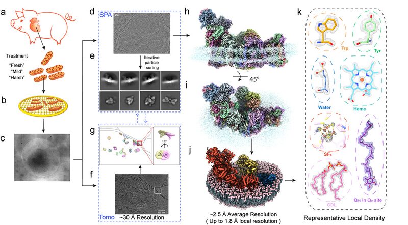
Traditionally, studies required purifying proteins from mitochondria before analysis, a process that could potentially damage protein activity. Leading the joint research team, Professor Zhu Jiapeng from Nanjing University of Chinese Medicine and Professor Zhang Kai from Yale University, have pioneered a new approach to overcome this challenge.
\"The in vitro purification method allows us to 'take pictures' of the mitochondrial proteins, but these images may differ from their real state. It is like a group of children playing on the playground. If I call one of the children to take a photo, they would likely act very prim. However, that is not what the child really looks like while playing,\" explained Professor Zhu.
The breakthrough lies in the ability to image intact mitochondria within a cellular environment, akin to taking realistic snapshots of children while they’re playing. The research team has achieved high-resolution imaging of mitochondrial proteins from cardiac origins at the level of intact organelles, reaching a resolution of up to 0.18 nanometers. This clarity allows for the observation of every atom within the protein structure.
These advancements pave the way for deeper investigations into the impacts of various mitochondrial diseases and pharmacological treatments by revealing reactive protein structures under physiological conditions within mitochondria. The study was published on May 29 in the journal Nature.
Reference(s):
cgtn.com