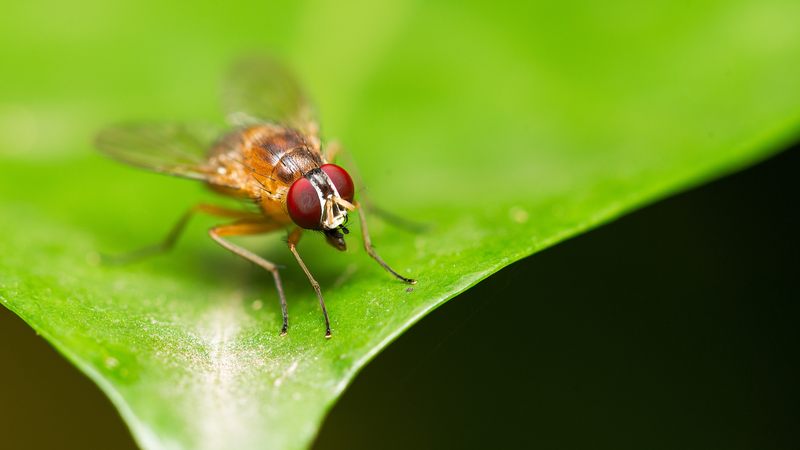
In a first-of-its-kind study, researchers at BGI Research in Hangzhou and the Southern University of Science and Technology in Shenzhen have crafted a groundbreaking 3D spatiotemporal multi-omics atlas that maps every single cell in the fruit fly throughout its entire life cycle—from embryo to adult.
By integrating data across transcriptomics, epigenomics and proteomics, the atlas offers a molecular time-lapse of development in vivid detail. The team tracked millions of cells as they transitioned through key stages—embryonic, larval, pupal and adult—uncovering dynamic gene networks and regulatory landscapes that drive growth, differentiation and metamorphosis.
A Molecular Time-Lapse
The project leveraged cutting-edge imaging and sequencing to capture 3D snapshots at dozens of time points. Each cell’s unique molecular signature was recorded, revealing how genes switch on and off as tissues form and specialize. This high-resolution map sheds light on critical windows where developmental defects may arise.
Why It Matters
Fruit flies (Drosophila melanogaster) are a powerhouse model for studying genetics and disease. With this atlas, scientists can now:
- Pinpoint stages when genetic mutations cause defects
- Trace the origins of developmental disorders at single-cell resolution
- Develop targeted treatments by understanding molecular drivers
The findings, published in Cell, open new avenues for research into birth defects, neurodevelopmental disorders and regenerative medicine. As a free resource for the global scientific community, the atlas exemplifies how collaborative efforts on the Chinese mainland can fuel discoveries with real-world impact.
Explore the data and join the conversation as we decode life’s blueprint—one cell at a time.
Reference(s):
cgtn.com